為納米技術研究市場提供光學圖像處理的CytoViva公司將一種新型光譜圖像技術(HSI)引入到**納米顯微鏡系統中����。這種技術的結合將幫助科學家在納米醫(yī)學和納米材料研究取得重大進展����。 在過去的幾年里,CytoViva的“納米顯微鏡系統"被很多****納米技術研究機構采納����,包括世界研究醫(yī)院�,約翰霍普金斯醫(yī)學院�、德州安德森癌癥中心、美國FDA�����、NASA����,NIH等。這一技術受到廣泛好評�,并獲得兩項RD100大獎,一項 Nano50 大獎���。

CytoViva?納米熒光高光譜顯微成像系統(HSI)�,可以同時提供納米材料及生物樣品的光譜分析和圖像數據��。該系統在可見-近紅外光譜范圍內(VNIR)進行數據采集�����,提供定量及定性分析數據���,對活體細胞和納米材料的熒光或非熒光成分適用�。
雙模式熒光顯微成像系統:支持熒光標記和非熒光標記物質同時觀察��;支持無機物質如:liposome�����,環(huán)碳納米管���,及其它納米粒子觀察和組織切片����、細胞的熒光/非熒光成像��;支持光譜分析等功能���。
CytoViva?是一種應用在生物醫(yī)學和納米技術研究領域的緊湊��,超高對比度���,超高分辨率,實時光學顯微鏡�����。該CytoViva?的光學照明系統是****的,取代了標準的顯微鏡聚光器�,并提供優(yōu)化的光學分辨率和檢測?����?茖W家們可以快速��,方便地觀察各種各樣的納米材料����。觀察到在傳統光學成像技術無法觀測到的,活細胞和**的細節(jié)���。通過將CytoViva雙模熒光系統��,研究人員還可以觀察到熒光標記的納米顆粒����,與無標記的**和活細胞的相互作用��。這種獨特的技術可以無需計算機的特殊處理��,只需兩種不同的照明方式和**的軟件程序���。
CytoViva?是一種**性的產品�,能夠創(chuàng)造新的熒光和高分辨率結合的光學顯微成像��,達到****的水平�����。這種獨特的系統使研究人員能夠實時的�����,高分辨率的同時觀察兩個熒光和非熒光樣品的結構�。擁有CytoViva?,研究人員現在就可以在自己的實驗室����,隨時動態(tài)觀察亞100nm的納米材料和生物活體標本的高分辨率成像。
CytoViva?將您現有的研究顯微鏡的性能提升到一個新的水平�。
實時觀察:
· 活細胞和納米材料的亞100納米分辨率
· 同時熒光和非熒光樣品結構
納米高光譜顯微成像系統:高光譜成像系統配合納米顯微成像系統,可以廣泛應用于量化細胞和組織中的納米材料�����。該系統捕獲掃描范圍內近紅外(400-1000nm)內每個像素的光譜信號�����;每一像素的完整光譜被CCD采集,光譜分辨率高達2nm�����,這些數據同時以光譜曲線和RGB圖像形式呈現��,每一個視野的詳細定量分析可以被執(zhí)行�,**的分析軟件可以提供的掃描材料的詳細光譜信息。適用于納米**輸送和納米毒理學領域���。 此高光譜分析方法既支持活細胞和納米材料的非熒光標記��,也支持熒光標記�����。
雖然這個系統是專門設計用于支持納米級的研究��,同時它也支持一個廣泛的�����,如細胞生物學和傳染性**領域的生物醫(yī)學研究�����。
Dr. Carlos Lopez-Estrano : University of Memphis College of Medicine
Dr. John Elliott: NIST
納米熒光高光譜成像
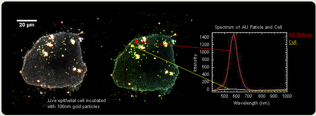
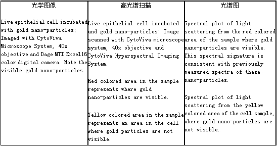
下面的圖片是CytoViva?系統在納米生物研究領域有代表性的一個典型應用����。此文章說明了CytoViva?系統對100nm的納米金顆粒�����,和一個活上皮細胞結合能力的量化����。
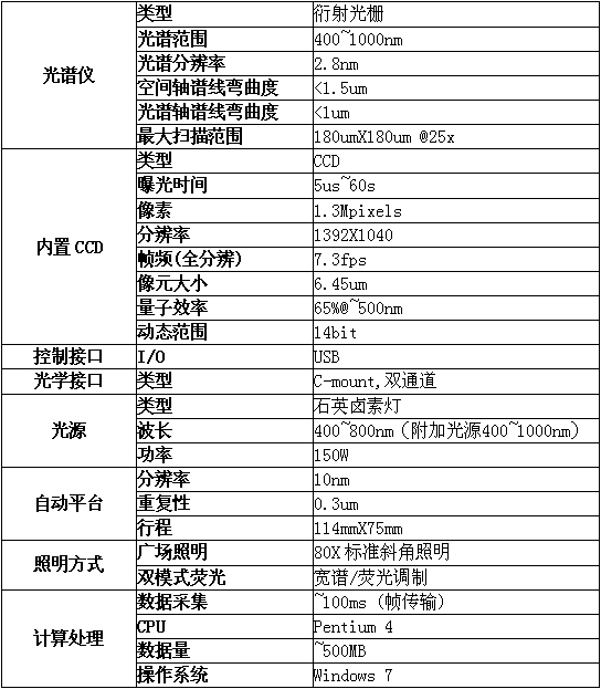
技術參數:
應用領域:
納米材料、納米醫(yī)藥�、納米**遞送、納米毒理學�、細胞生物學、病理學�����、病毒學����、植物學......
英文介紹
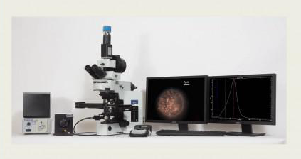
The CytoViva® Microscope System
Specifically designed to support research in nanotechnology and infectious disease, CytoViva employs a patented (US patents No. 7,542,203, 7,564,623) darkfield-based optical illumination system. This structured illumination technology replaces the standard condenser on a research grade microscope. By improving the alignment and focus of darkfield or oblique angle illumination, the technology enhances signal-to-noise of nanoscale samples up to seven times over standard darkfield optics. This enables scientists to optically observe a wide range of nanoscale materials quickly and easily in solution, live cells, tissue and materials based matrices. In addition, non-fluorescent live cells and pathogens can be easily observed at a level of detail not possible with traditional optical imaging techniques such as phase contrast or differential interference contrast.
When using the CytoViva Dual Mode Fluorescence system, researchers can also observe the interactions between fluorescently labeled nano-particles or bacteria and live unlabeled cells. This unique capability can eliminate the need to create computer enhanced overlay images which require two different illumination methods and advanced software programs. Finally, when combined with CytoViva’s Hyperspectral Imaging capability this high signal-to-noise microscopy method enables researchers to spectrally characterize and map nanoscale samples in a wide range of environments.
用戶評價:
J. Paul Robinson, PhD, Director of Purdue University's Cytometry Laboratory, Professor of Basic Medical Sciences and Biomedical Engineering, Purdue University comments...
""It's like not knowing you need glasses. You don't know what you can't see. Then someone hands you a pair-and the world is clear with amazing detail.""
John A. Smith, MD, PhD, MMM, Divisional Director, Department of Pathology, University of Alabama-Birmingham says it well ...
""Looking through CytoViva, you are face to face with living biology. You see into the world of cell biology
that you didn't know existed. You are visualizing the future of underlying biological processes as it merg
es with the present.""
Dr. Elaine Coleman, Associate Professor, Auburn University, Department of Anatomy, Physiology and Pharmacology comments...
""Its capabilities in cell culture research are astounding. I have purchased a unit for my future research because its capabilities are unique for observing live cell cultures.""
部分用戶:
National Institute for Occupational Health
US Food and Drug Administration
Finnish Institute for Occupational Health
National Institute of Health Sciences Japan
Melbourne Center for Nanofabrication
US Army Corp of Engineers
Lawrence Berkeley National Labs
IIT Madras
Rice University
Fraunhofer Institute
Adolph Merkle Institute
University of Sao Paulo
Health Canada
Stanford University
University of Bonn
Georgia Tech University
Duke University
University of Montreal
Wright Patterson AFB
MD Anderson Cancer Center
Center for Nanoscale Science and Engineering
文獻:
有超過300篇用CytoViva?納米熒光高光譜顯微成像系統發(fā)表的第三方文獻����,這是*近發(fā)表的部分文獻:
Hyperspectral imaging for cellular iron mapping in the in vitro model of Parkinson's disease
Eung Seok Oh, Chaejeong Heo, Ji Seon Kim, Minah Suh, Young Hee Lee, Jong-Min Kim
Journal of Biomedical Optics | Volume 19 | Issue 5 | Special Section on Nanobio-Based Optical Sensing and Imaging 2014
Optical hyperspectral investigation of the J-pole and Vee antenna families
Timothy D. James, Timothy J. Davis, and Ann Roberts
Optics Express, Vol. 22, Issue 2, pp. 1336-1341 (2014)
In Vitro Identification of Gold Nanorods through Hyperspectral Imaging
Bradley M. Stacy, Kristen K. Comfort, Donald A. Comfort, Saber M. Hussain
Plasmonics June 2013, Volume 8, Issue 2, pp 1235-1240
Hyperspectral enhanced dark field microscopy for imaging blood cells
Giulia Sacco Verebes, Michele Melchiorre2, Adianez Garcia-Leis, Carla Ferreri, Carla Marzetti,
Armida Torreggiani
Journal of Biophotonics Volume 6, Issue 11-12, pages 960–967, December 2013